
Short answer: Start with whether fixation or permeabilization will follow. PI or 7-AAD may suit surface flow cytometry on unfixed samples when the detection channel fits the panel. For workflows requiring fixation, permeabilization and intracellular staining, consider a validated fixable viability dye: stain and wash before fixation, then proceed with the remaining steps.
A familiar viability stain may work well for surface-antigen analysis. Adding intracellular antigens, cytokines or transcription factors introduces fixation, permeabilization and additional washes, so the original dye and staining sequence need to be reviewed.
Write down the experimental workflow before choosing a color. The aim is to retain suitable cell populations for analysis and reduce interference such as nonspecific antibody binding by membrane-compromised cells.
When should you consider PI, 7-AAD or a fixable dye?
PI and 7-AAD are widely used nucleic-acid-binding viability dyes. They have limited access through intact cell membranes but can enter membrane-compromised cells and bind nucleic acids. They are commonly used to exclude dead cells from unfixed samples.
PI can bind DNA and RNA, whereas 7-AAD preferentially binds DNA. Their emission spectra differ, so assess the detection filters, existing panel and compensation settings separately.
Amine-reactive fixable dyes such as Cell-Life form covalent labels with accessible protein amines. They can record membrane-integrity differences before fixation and support subsequent analysis with a validated fixation and permeabilization workflow.
| Candidate type | When to consider it | Fixation and permeabilization checks |
|---|---|---|
| PI | Surface flow cytometry on unfixed cells, with a suitable detector | Do not treat it as the default way to recover pre-treatment viability after fixation or permeabilization; follow the reagent instructions for pre-acquisition staining |
| 7-AAD | Surface flow cytometry on unfixed cells, with a compatible channel in the existing panel | Check downstream processing; it is not directly interchangeable with an amine-reactive fixable dye |
| Amine-reactive fixable viability dye, such as Cell-Life | Subsequent fixation, permeabilization or intracellular staining; also validated unfixed workflows | Stain before fixation and permeabilization, remove unreacted dye, and validate live/dead separation through the complete workflow |
Common PI/7-AAD viability-staining procedures retain dye in the acquisition buffer. Follow the instructions for the specific reagent when deciding whether to wash after staining; do not simply apply the Cell-Life step of removing unreacted dye.
PI and 7-AAD may have other nucleic-acid-staining uses in fixed samples. The question here is whether staining identifies the viability state before processing. Fluorescence after fixation does not by itself demonstrate that this earlier information has been retained.
How does a fixable dye distinguish intact and compromised cells?
Cell-Life is an amine-reactive dye. In cells with intact membranes, it mainly encounters reactive amines on the cell surface, typically producing a lower signal. In membrane-compromised cells, it can also access more intracellular proteins, typically producing a stronger signal.
Some fluorescence from live cells is therefore not necessarily a failed stain. Assess reagent amount, live/dead controls and population separation rather than requiring the live-cell signal to be zero.
This live/dead distinction is based on membrane integrity at the time of staining. The readout alone does not identify early apoptosis, and low staining does not establish normal metabolic function.
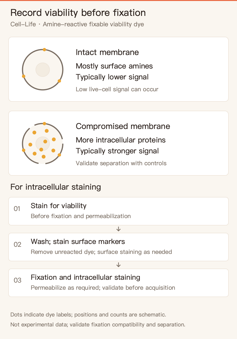
Figure 1. Principle and sequence of amine-reactive viability staining. This qualitative diagram does not specify molecular counts, fluorescence ratios or experimental data; validate population separation with appropriate controls.
Fixability supports suitable downstream processing. Compatibility with a particular fixative, permeabilization system or storage period still requires verification across the complete workflow.
Where should viability staining fit into the workflow?
In the Cell-Life reference flow cytometry procedure, collect and wash cells, stain for viability in a suitable buffer, and remove unreacted dye. Then perform surface-antigen staining, fixation, permeabilization and intracellular staining as required by the assay. Finally, resuspend the cells for acquisition.
To preserve information about the original membrane-integrity state, stain before fixation or permeabilization. Adding a viability dye to cells that have already been fixed or permeabilized cannot reliably reconstruct the earlier live/dead state.
Check the staining buffer separately. The Cell-Life flow cytometry instructions specify PBS for washing and resuspension at this stage and warn against using BSA- or serum-containing FACS buffer because proteins may interfere with staining. After staining and washing, use the buffers specified for subsequent steps. This is not a requirement to keep every later step protein-free.
The reference flow cytometry conditions were developed with Jurkat cells. Reassess working dye amount and incubation conditions for primary cells, other cell lines or different cell numbers. Use the microscopy and flow cytometry instructions for their respective applications.
How should you select a Cell-Life color for a multicolor panel?
Check the instrument's laser lines, filters and channels already assigned to antibodies, then identify a suitable remaining detection position. Qihe offers different Cell-Life spectral options; the following three provide starting points for review:
| Product | Common laser line listed in the instructions | Further selection checks |
|---|---|---|
| Cell-Life Dye 405/450 | 405 nm | Whether other violet-laser markers already occupy the corresponding detector |
| Cell-Life Dye 488/520 | 488 nm | Spectral overlap, spreading and population-separation effects involving existing green markers |
| Cell-Life Dye 633/780 | 633–640 nm | A detector covering emission near 780 nm and compatibility with other panel colors |
The numbers retained in product names and the laser lines in the table serve different purposes. Do not confuse laser wavelength, emission maximum and filter center wavelength. Color names alone also cannot establish whether two labels can be combined.
Conventional flow cytometry requires assessment of compensation and spillover spreading; spectral cytometry requires appropriate reference spectra and evaluation of unmixing. Strong dead-cell staining may affect other channels, so evaluate performance in the complete panel.
What should you check if staining is too strong, too weak or poorly separated?
Start with cell preparation. Excessive handling, repeated centrifugation or unsuitable dissociation conditions may affect membrane integrity. Interpret the viability readout in the context of sample processing.
Next review the staining solution and reagent amount. Check for residual protein-containing culture medium, correct dye preparation and a suitable relationship between cell number and working dye amount. Titrate for clear live/dead separation rather than maximizing fluorescence in every cell.
Then review controls and acquisition settings. An unstained control helps assess autofluorescence. Suitable live and membrane-compromised cell controls help locate the populations. Multicolor assays also need a single-stain control compatible with the dye for compensation or unmixing. Controls for fixed or permeabilized assays should receive the same treatment; compare separation before and after processing when needed.
For product selection or a quote, send the instrument model, laser and filter configuration, existing staining panel, cell type and fixation/permeabilization plan to the Qihe technical team. These details help identify candidate Cell-Life options and validation needs. Browse the assay kit catalog for additional spectral options.
Frequently asked questions
Can I stain already-fixed cells to identify which cells were originally alive?
Do not rely on a viability stain at that point to reconstruct the original live/dead state. Fixation and permeabilization change membrane integrity. Plan appropriate staining and controls before processing when that information is required.
Live cells have some Cell-Life fluorescence. Should I add more dye?
Do not increase dye based on that observation alone. Live-cell surfaces also contain reactive amines, so low surface staining can occur. Assess live/dead separation first, then review reagent amount, washing and cell handling.
Does choosing a fixable dye remove the need for live/dead controls?
No. Controls establish population positions for the actual sample, instrument and complete workflow. Multicolor assays still need single-stain controls to assess the required compensation or unmixing settings.
This article provides reagent-selection guidance for in vitro research. Follow the applicable product instructions for preparation, use and storage, and validate performance in the intended sample and complete assay workflow.