
Short answer: Confirm that the antibody is biotinylated, then choose fluorescent streptavidin to match the instrument's lasers, available detectors and complete staining panel. In conventional flow cytometry, APC and AF647 often use similar far-red detection channels; different names do not establish independent colors.
If you already have a suitable biotinylated antibody, choosing between streptavidin-PE, streptavidin-APC and AF647-Streptavidin is a practical part of building a flow cytometry assay.
All three use streptavidin to recognize biotin. The choice concerns which fluorescent reporter suits the instrument, sample and existing panel, and how its performance will be assessed in the intended assay.
How a biotinylated antibody becomes a fluorescent readout
A biotinylated antibody carries a biotin tag but does not necessarily carry a fluorophore. After the antibody binds its target antigen, fluorescent streptavidin can bind the antibody's biotin and provide a signal in a matched detector.
The antibody recognizes the target, biotin supplies the attachment site, and fluorescent streptavidin supplies the reporter. Streptavidin recognizes biotin rather than the species of the primary antibody. It therefore cannot simply replace a species-specific secondary antibody for an unlabeled primary.
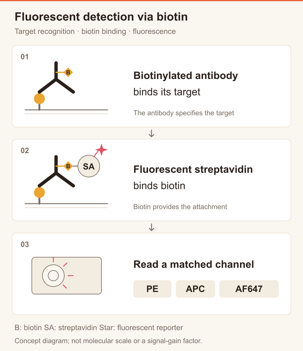
Figure 1. Detection with a biotinylated antibody and fluorescent streptavidin. This concept diagram does not specify molecular scale, binding-site counts or signal amplification factors.
The same biotinylated antibody can be evaluated with different streptavidin colors, providing flexibility in channel assignment. After changing colors, reassess reagent amount, background and detection settings rather than assuming that the previous conditions transfer unchanged.
Start with the instrument before comparing PE, APC and AF647
Check the laser lines, optical filters and channels already occupied by other markers. Labels such as PE or APC in the acquisition software are starting points; verify the actual optical configuration.
| Candidate product | Common excitation configuration | Main selection checks |
|---|---|---|
| R-PE-Streptavidin | 488 nm blue or 561 nm yellow-green laser | A suitable PE detector; spillover and spreading effects within the existing panel |
| APC-Streptavidin | 633–640 nm red laser | An available far-red detector; sample background and other red-laser markers |
| AF647-Streptavidin | 633–640 nm red laser | Spectral compatibility, overlap with APC-like colors and the measured signal-to-background relationship |
These are common instrument excitation configurations, not the excitation maxima of the products. They do not imply equal detection efficiency. Confirm the spectrum of the purchased reagent against the instrument configuration.
APC and AF647 often use similar far-red detectors in conventional flow cytometry. If APC already occupies that channel, AF647 should not normally be added as an independent color without redesigning the panel. Spectral cytometry also requires assessment of reference spectra and actual unmixing performance; names alone do not establish compatibility.
What else matters after choosing a color?
R-PE and APC are phycobiliprotein reporters, while AF647 is an organic fluorescent dye. Every conjugate also contains streptavidin protein. AF647-Streptavidin as a complete reagent is therefore not a small-molecule dye, and properties of an isolated fluorophore do not establish compatibility of the whole conjugate.
Fixation, permeabilization and planned storage affect selection and validation. Check the instructions for the specific product. Qihe's documentation specifies that its R-PE and APC conjugates should not be frozen.
For a weak target signal, the product name or color alone cannot determine the final choice. Compare candidates using the same antibody, comparable samples and suitable detection conditions. Assess positive–negative separation, background and effects on other channels. A higher fluorescence reading does not necessarily provide better resolution.
What should you check when signal is weak or background is high?
First check the detection chain: Is the antibody biotinylated? Does it bind the target in this sample? Does the streptavidin reagent carry the intended fluorescent reporter? Are the laser and detector settings appropriate?
If the antibody was biotinylated in-house, review purification. Residual free biotin can occupy streptavidin binding sites and reduce effective detection of the biotinylated antibody. Use appropriate purification for the labeling method rather than treating extra streptavidin as a general remedy.
For high background, unstained samples and a control receiving fluorescent streptavidin without the biotinylated primary can help identify the source. In fixed or permeabilized samples, consider endogenous biotin as well as cell condition, nonspecific binding and reagent amount. Multicolor assays also require suitable single-color controls for compensation or spectral unmixing. Include fluorescence-minus-one (FMO) controls when needed to assess weak positive populations.
Evaluate reagent concentration for the actual sample and panel, balancing positive separation against negative background. A dilution used successfully elsewhere is not a universal setting for every antibody and sample.
What should you provide when requesting a quote?
Share the instrument model, laser lines and filter configuration; antibody identity and biotinylation status; sample type and fixation or permeabilization requirements; and the existing staining panel and current problem.
Qihe offers fluorescent streptavidin conjugates including R-PE, APC and AF647, with additional options for other detection channels. Browse the Streptavidin conjugate range, or send these details to the Qihe technical team to discuss candidate products, pack sizes and validation needs.
Frequently asked questions
Can fluorescent streptavidin directly detect an antibody that is not biotinylated?
Do not assume this is a matched detection pair. Streptavidin recognizes biotin, so an accessible biotin tag is needed, or a different detection route appropriate for the antibody must be selected.
Is changing the reagent name enough when switching from APC to AF647?
Recheck the spectrum, instrument settings, reagent amount and controls. The reagents may use similar detectors, but their signal, background and compensation or unmixing requirements still need validation.
Can two streptavidin colors distinguish two biotinylated antibodies?
If two biotinylated antibodies and two streptavidin colors are added together in the same assay, both streptavidin reagents recognize biotin. Color alone cannot assign each reporter to a particular antibody. Use a separately designed, distinguishable labeling strategy for independent targets.
This article provides reagent-selection guidance for in vitro research. Follow the applicable product instructions and validate performance in the intended sample and assay.